Sample Preparation
Homogenization
Heating and Mixing
Electrophoresis and Blotting
Polyacrylamide Gel Electrophoresis
Agarose Gel Electrophoresis
Western Blotting
Power Supplies
PCR & qPCR Thermal Cycler
Thermal Cycler (PCR)
Real-time Thermal Cycler (qPCR)
PCR Workstations & Cabinets
UVP BioImaging Systems
Molecular Spectroscopy
Lab Equipment
Ultraviolet Products
Hybridization Ovens
UVP Incubator
UV Crosslinkers
UVP Benchtop Transilluminators
Thermal Mixers
Electrophoresis & Blotting
Thermostats
View All
Fume hood
Laminar Airflow
Biosafety Cabinet
Autoclave
Centrifuge
pH Meter
Shaker & Mixer
Orbital Shaking Incubator
BOD Incubator
Heating Oven
Water Purification System
Aermax - Air Purification
Medical Oxygen Concetrators
Hygiene Solution
-150°C Cryogenic Freezer
-86°C Ultra Low Temp Freezer
-40°C Low Temp Freezer
-18 ~ -25°C Biomedical Freezer
-20°C Biomedical Freezer
4° ± 1°C Blood Bank Refrigerators
2~8°C Pharma Refrigerators
2~8°C ICE Lined Refrigerators
-25°C ~ + 4°C Mobile Freezer/Collers
20~24°C Blood Platelet Incubators
Ice Machines
Coldrooms
Mortuary Chambers
Zone electrophoresis and moving boundary electrophoresis are the main types of electrophoresis.
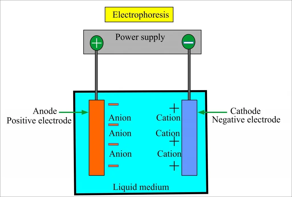
Zone electrophoresis involves the migration of charged molecules in a solution with the supporting medium. The separated components are formed like discrete zones in the supporting medium. Buffer solution saturates the supporting medium and a small volume of sample is applied as a narrow band.
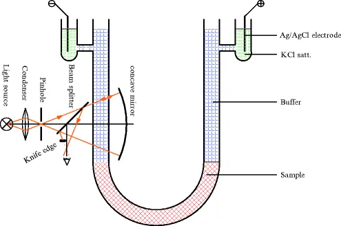
Moving boundary electrophoresis involves the migration of charged molecules in a free moving solution, without the presence of a supporting medium.
Paper electrophoresis, thin-layer electrophoresis, gel electrophoresis, and cellulose acetate electrophoresis are the types of zone electrophoresis. Moving boundary electrophoresis is classified into capillary electrophoresis, isoelectric focusing, isotachophoresis, and immunoelectrophoresis.
It involves the carrying out electrophoresis in a thin layer of silica or alumina for the separation of amino acids.
Cellulose acetate electrophoresis is a modification of paper electrophoresis. Cellulose acetate is prepared by treating acetic acid with cotton, having sulfuric acid as a catalyst. The migration of the sample takes place in the buffer on the cellulose surface plate based on the charge.
The support medium is filter paper, which can be Whatman no. 1 and Whatman no. 3. The width of the paper used in the process is 5 cm. The separation of molecules will take place for up to 16-18 hours. There are two troughs in the apparatus, containing buffer solution, through which electric current is passed. A Homogeneous group of molecules migrates as a separate band. Using fixative like methanol or acetone, separated molecules are fixed to solid support.
The separation of molecules is performed through the molecular sieving technique, which is based on the substances’ molecular sieve. Gel, a colloid, acts as a molecular sieve. It should be electrically neutral. Agarose, polyacrylamide, starch, and Sephadex gels are used as gel materials. The gel allows smaller molecules to pass through, at the same time, obstructing the movement of larger molecules.
Agarose is a purified uncharged polysaccharide, extracted from seaweeds. It has repeating units of 3,6-anhydro-L-galactose. On adding agarose to a boiling liquid, it remains as a liquid up to 40°C. On lowering further, it changes into a gel state. By manipulating the concentration of agarose in the gel, the pore size can be adjusted. The agarose gel is formed by weak hydrophobic and hydrogen bonds, which make the compound fragile.
It is prepared by boiling granular starch suspension in a buffer to provide a colloid. On cooling, colloid turns into a semi-solid gel, due to branched chains of amylopectin. Petroleum jelly is used for the prevention of shrinking and swelling.
Polyacrylamide gel is prepared by polymerization of acrylamide in the presence of crosslinking agent methylene-bis-acrylamide. Covalent cross-link holds together acrylamide monomers. It is tougher than agarose gel and is relatively inert, thermostable, strong and transparent. It is prepared in numerous pore sizes and is uncharged. Protein molecules are separated on the basis of molecular size and charge to mass ratio.
It is run in non-denaturing conditions leading to the preservation of the analyte's natural structure. Separation is done on the basis of size, shape and charge of molecules. It is the original model and it is called native polyacrylamide gel electrophoresis (PAGE). It is useful in the purification or separation of a mixture of proteins.
It is a commonly used method for the determination of the molecular weight of proteins. The separation is based on molecular weight and electrophoretic mobility. Detergent SDS is added to PAGE, so it is called SDS PAGE. SDS provided a constant mass-charge ratio by coating with proteins. Migration of proteins happens in the order of increasing molecular sizes or weights. In proportion to the protein length, SDS confers a negative charge, thereby destroying non-sulfide-lined tertiary and all secondary structures. Hence, molecules will have a net negative charge with a broad pH range. On binding with SDS, proteins become charge densities and the proteins separate on the basis of mass.
Isotachophoresis is meant by moving at the same velocity. It works on the development of potential gradients. Leading and trailing electrolytes are used. The leading electrolyte has more mobility than the fastest sample and the trailing electrolyte has lesser mobility than the slowest sample. After the voltage is applied, the leading electrolyte migrates quickly towards the corresponding electrode, with the sample ions following immediately, creating zones in the order of mobilities.
Isoelectric focussing works on the development of pH gradients. Each protein is migrated to an area of specific pH. The pH of the gradient equals the pH of the protein, leading to the formation of well-defined and sharp protein bands. According to the isoelectric point, the distance of migration happens. The net charge of the protein becomes zero at the stoppage of migration and zwitterion is formed. When the solution’s pH is below the pI of protein, protein is positively charged and migrates towards the cathode. When the solution’s pH is above the pI of protein, protein becomes negatively charged and migrates towards the anode. The electrolyte is an ampholyte, having carboxylic acid and aliphatic amino groups, which contain a range of isoelectric points.
Capillary electrophoresis involves filling capillaries with buffers and application of high voltages to obtain separation of molecules. The capillaries are narrow, with an internal diameter of 25 - 100 nm. The capillaries are filled with a gel eliminating the electro-osmotic flow and the process is run like conventional electrophoresis, allowing greater resolution, higher sensitivity, and online detection. Electrodes are also inserted in the reservoirs for the completion of the circuit.
When an electric current is applied to a slide filled with a layer of gel, a mixture of antigens present in the wells is separated into individual antigens based on the size and charge. Then, the separated molecules are allowed to react with specific antisera placed in troughs, placed parallel to electrophoretic migration and diffusion takes place. Antisera in the troughs migrate towards antigens, react with them, forming separate precipitin lines, which is indicative of the reaction of individual proteins with antibodies.
Conclusion
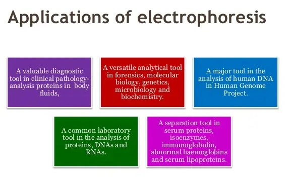
We have seen the different types of electrophoresis. Each type of electrophoresis has its own characteristic and specific. The cost of each type of technique also differs. Depending on the needed sensitivity of the experiment and the cost involved, the type of electrophoresis is chosen. In addition, each electrophoresis has its own pros and cons and applications too.