Sample Preparation
Homogenization
Heating and Mixing
Electrophoresis and Blotting
Polyacrylamide Gel Electrophoresis
Agarose Gel Electrophoresis
Western Blotting
Power Supplies
PCR & qPCR Thermal Cycler
Thermal Cycler (PCR)
Real-time Thermal Cycler (qPCR)
PCR Workstations & Cabinets
UVP BioImaging Systems
Molecular Spectroscopy
Lab Equipment
Ultraviolet Products
Hybridization Ovens
UVP Incubator
UV Crosslinkers
UVP Benchtop Transilluminators
Thermal Mixers
Electrophoresis & Blotting
Thermostats
View All
Fume hood
Laminar Airflow
Biosafety Cabinet
Autoclave
Centrifuge
pH Meter
Shaker & Mixer
Orbital Shaking Incubator
BOD Incubator
Heating Oven
Water Purification System
Aermax - Air Purification
Medical Oxygen Concetrators
Hygiene Solution
-150°C Cryogenic Freezer
-86°C Ultra Low Temp Freezer
-40°C Low Temp Freezer
-18 ~ -25°C Biomedical Freezer
-20°C Biomedical Freezer
4° ± 1°C Blood Bank Refrigerators
2~8°C Pharma Refrigerators
2~8°C ICE Lined Refrigerators
-25°C ~ + 4°C Mobile Freezer/Collers
20~24°C Blood Platelet Incubators
Ice Machines
Coldrooms
Mortuary Chambers
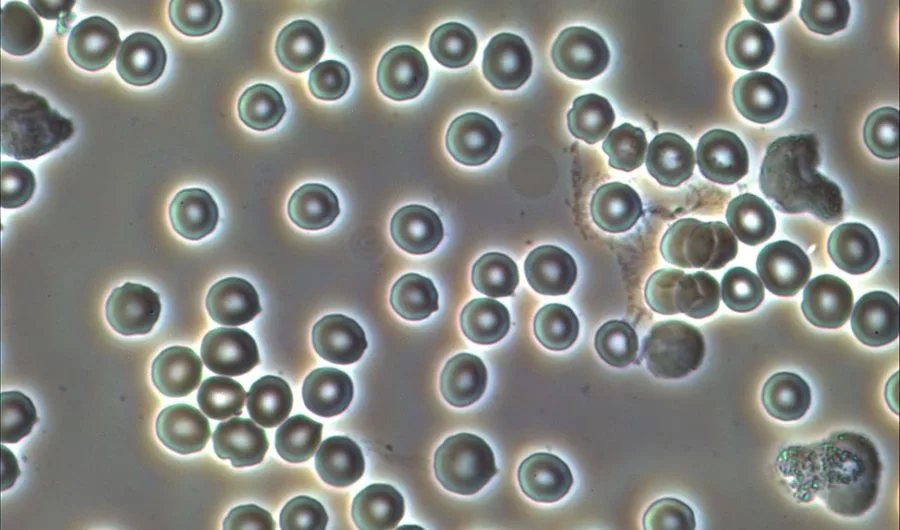
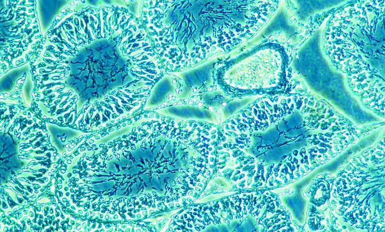
Microscopy techniques are widely used in laboratories for observing small particles, cells, etc. In this case, three-dimensional live imaging has become the requirement of research labs. This live imaging is a fundamental part of cell development observations and neural biology applications. To obtain such structural details in the cell cultures, some precise Spatio-temporal manipulations are done to the biological specimens to give out good results. For example, the phase-contrast microscopy technique can use the process of reflection and diffraction to produce high contrast images. Here, the specimens used are of thin membranes so that light rays diffract from them. Hence, such manipulations lead to a better vision of the biological samples.
In the technique of pulsed illumination phase-contrast microscopy, the optical design of the concerned microscope is integrated with the short infrared laser pulses. One more addition to these is the Spatio-temporal three-dimensional light-sheet imaging technique. All these three help in executing the pulsed illumination phase-contrast microscopy for a biological sample. The infrared laser pulses induce the Spatio-temporal optical manipulations which tend to bring out the high pulsed illuminated images. Demonstrations on Drosophila melanogaster and zebrafish model systems have given a lot more insights into the pulsed illumination phase microscopy techniques than any other microscopy technique.
Pulsed illumination is the breakthrough discovery in the history of microscopy technologies. The optical arrangement of pulsed illumination has a close resemblance with the normal optical arrangement of an inverted phase microscope. The inverted microscope has a normal sample space for the placement of biological specimens. Laser light pulses are also placed in the concerned phase contrast microscope for sourcing the pulse’s light rays. All these arrangements are called optical arrangements. Here, the light rays allow a pulse repetition frequency to generate the images of all biological specimens in high contrast nature. However, the generated pulse rates can be reduced easily by the researcher. Pulsed illumination phase microscopy is called a cost-intensive device which may reduce the stability of laser light used here. In such cases, the perfect illumination cannot be achieved.
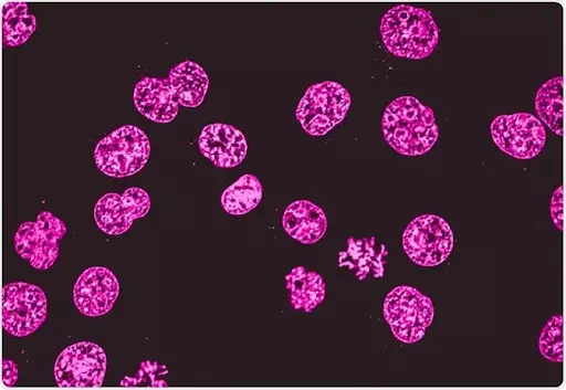
The pulsed illumination technique can be used in the cell culture microscope, tissue culture microscope, and culture microscope for wide observations. This type of optical arrangement is used by fluorescence microscopes also. These microscopes use particular confocal or light-sheet microscopes for perfect viewing. In these types of applications, pulse lasers are used for exciting dyes, like fluorophores or luminophores. These dyes then produce the afterglow followed by the prior excitation.
Infrared rays are the principal source for laser pulse illumination in phase-contrast microscopy and inverted phase microscope. The rays are capable of exciting the labeling dyes on the biological specimens which give out the illumination.
Following are the several benefits of using the IR rays for illumination :
In phase-contrast microscopy, the pulsed illumination technique is used by arranging the laser light source along with dyes. Some manipulations are also done with biological specimens for generating perfect illuminated images. Here, the laser light source may emit light of multiple colors or maybe a supercontinuum laser or a Raman comb laser. These are the name given to the laser lights emitted by the source. The laser light may include a pump light source and a non-linear fiber for illumination. The non-linear fiber is defined as the nonlinear optical effects, like self-phase modulation of the emitted laser pulses on the specimens. Through the laser light pump or light source in an inverted phase-contrast microscope, the laser light is emitted with at least one laser pulse. Here, the optical beam is the same as the optical path which propagates through electromagnetic waves of light or electromagnetic wave packets of light.
The pulsed illumination process uses the optical path in a rectilinear path and can be changed by optical elements, like prims, mirrors, gratings, etc. It is seen that the optical beam path is determined essentially by the laser light source used in pulsed illumination phase-contrast microscopy. In a normal microscope, the trinocular head may also possess the laser light source for emitting the laser light rays. In the pulsed illumination process, the transmission state of laser lights is understood as an adjustment or mode of the optical arrangement. Here, each of the transmission states is due to a respective emission spectrum of light rays. The rays are the characterization in this transmission spectra that exhibits different transmission states of linear laser rays. From the light source, the pulsed beams of light enter the sample stage where the specimen is placed. Some traditional light rays are so identical, that they can only be defined by different wavelength ranges. All this type of arrangement is called the optical arrangement for the pulsed illumination process for phase-contrast microscopy.
With the above arrangement, the laser clock is fitted with synchronization with laser light beams. This ensures that the laser pulse transmits with response to the emission required. This remains unchanged at least the next lighting cycle after the one complete cycle of illumination. The researchers can also use the lightning clocks for ensuring the transmission of laser beams to the biological specimens. Between the two lightning cycles, no transmission of light happens. With all this arrangement, pulsed illumination can be executed. It is very important to learn all components of pulsed illumination to obtain phase contrast images of biological specimens from the camera for a trinocular microscope.
Moreover, the infrared rays can penetrate deep into the cells and tissues to give out the perfect images. This is one more reason to use infrared rays as a laser beam of lights in pulsed illumination. Many biological specimens have deep layers and the normal light rays cannot penetrate them. This obstacle is overcome by using laser beams made up of infrared light. Many best trinocular microscopes also have room for laser beams for the illumination process. Scanning and fluorescent microscopes can also be employed with laser light beams for proper illuminations.
Conclusion
It is seen that phase-contrast microscopy is used for generating high contrast images of all biological samples. With the optical arrangement of laser beams, we can get the pulsed illuminated images of many biological specimens with detailed structures. Upright and inverted microscopes can also use the laser beam arrangement for proper illumination. Researchers and scientists can check the inverted microscope price online as well as offline medium.